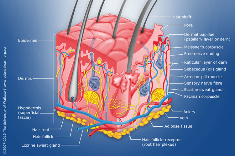
Diagram of human skin structure — Science Learning Hub
Dermis. Definition. Fibrous and elastic tissue, provides strength and elasticity to the skin and supports the epidermis, home to hair follicles, glands, nerves etc. Location. Term. Papillary Layer. Definition. Upper dermal layer, provides the epidermis with nutrients and regulates body temperature. Location.

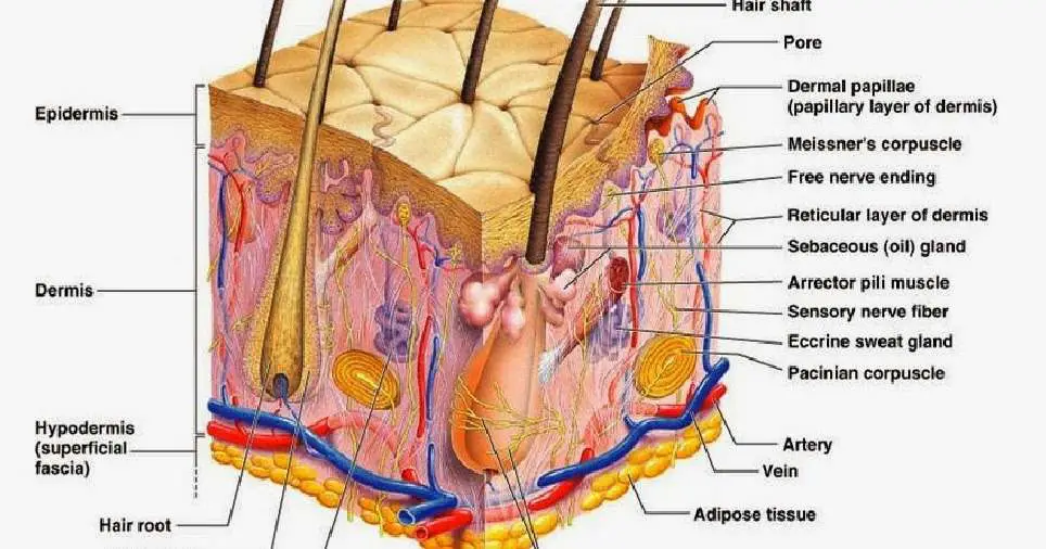
Cross section anatomy of skin with labels on white background
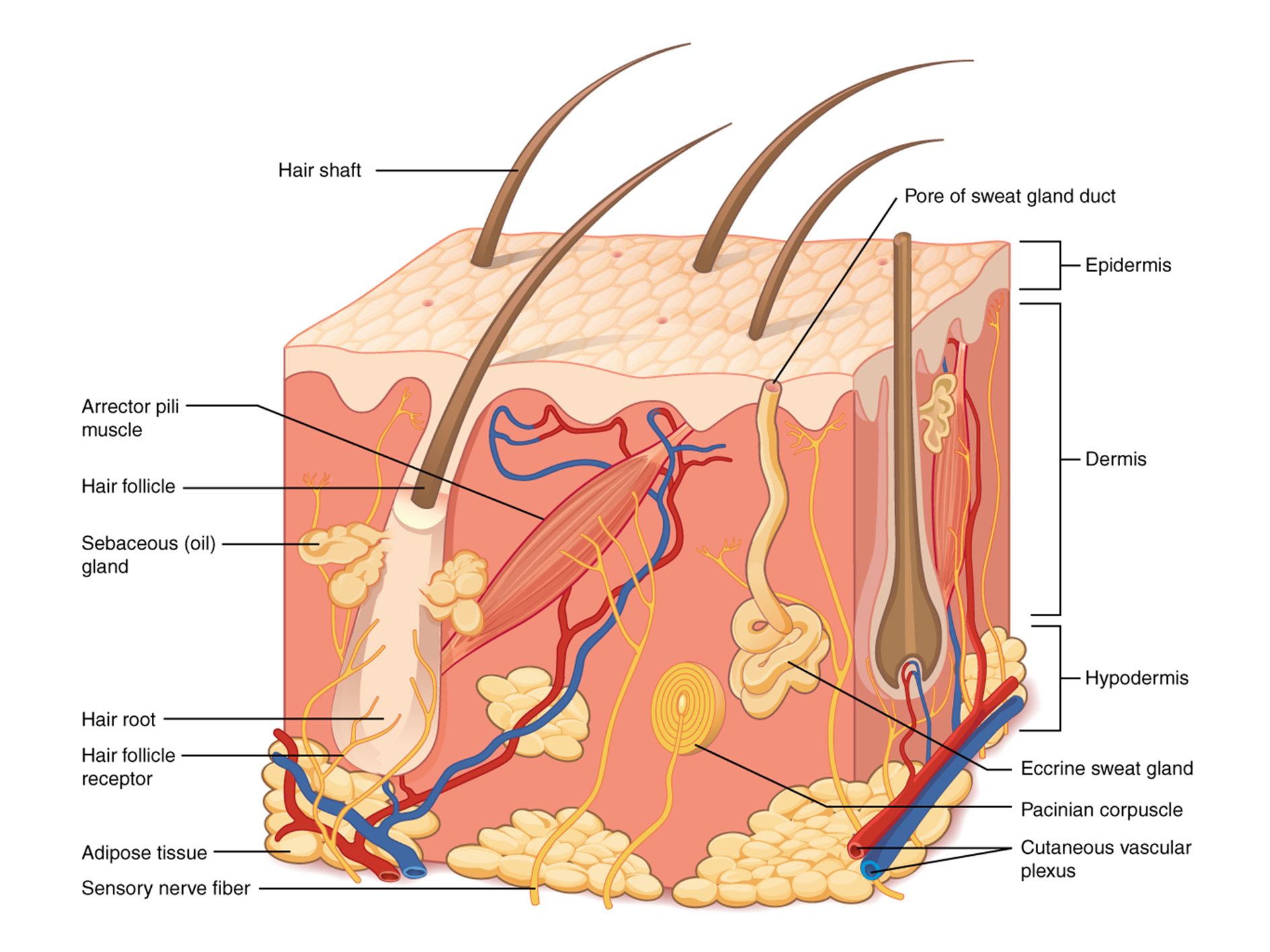
Diagram of human skin structure. Image. Add to collection. Rights: The University of Waikato Te Whare Wānanga o Waikato Published 1 February 2011 Size: 100 KB Referencing Hub media. The epidermis is a tough coating formed from overlapping layers of dead skin cells.

Human skin diagram Anatomy and Physiology in 2018 Pinterest Skin
Stratum Basale. The stratum basale (also called the stratum germinativum) is the deepest epidermal layer and attaches the epidermis to the basal lamina, below which lie the layers of the dermis. The cells in the stratum basale bond to the dermis via intertwining collagen fibers, referred to as the basement membrane. A finger-like projection, or fold, known as the dermal papilla (plural.
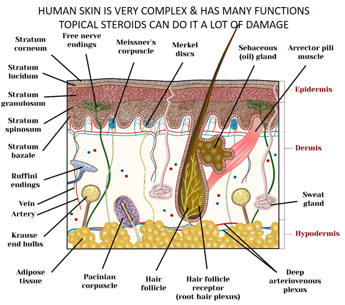
Skin topical steroid withdrawal with wheatgrass extract A
'Skin Diagram || How to draw and label the parts of skin' is demonstrated in this video tutorial step by step.The sense of touch had received supreme importa.

Science Homework, Skin Drawing, Human Body Anatomy, Sense Of Touch
Signs of skin infections like red streaks or yellow discharge. Unexplained skin rash or skin condition. A note from Cleveland Clinic. As the body's largest organ, your skin plays a vital role in protecting your body from germs and the elements. It keeps your body at a comfortable temperature, and nerves beneath the skin provide the sense of.

Skin Structure infographic LifeMap Discovery
The skin has a surface area of between 16.1-21.5 sq ft. for an adult human. The thickness of the skin differs over all parts of the body, and between men and women and the young and the old. For example, the skin on the forearm which is on average 1.3 mm in the human male and 1.26 mm in the human female.
Skin diagram to label Labelled diagram
1. The outermost layer of the skin is: the dermis / the epidermis / fat layer. 2. Which is the thickest layer: the dermis / the epidermis? 3. Add the following labels to the diagram of the skin shown below: Epidermis, dermis, fat cells, hair shaft, hair follicle, hair erector muscle, sweat gland, pore of sweat gland, sebaceous gland, blood.

Skin Structure (labelled), illustration Stock Image C043/4873
The skin is by far the largest organ of the human body, weighing about 10 pounds (4.5 kg) and measuring about 20 square feet (2 square meters) in surface area. It forms the outer covering for the entire body and protects the internal tissues from the external environment. The skin consists of two distinct layers: the epidermis and the dermis.
Skin diagram labeled
Labeled diagram of the skin. So what's the idea? Spend some time analyzing the skin diagram labeled above. Try to memorize the appearance and location of each structure. Learning the function of each structure will accelerate your ability to memorize, so be sure to check out our detailed article on The Integumentary System parts and functions.
Human Skin Structure, Useful Functions, and Interesting Facts Owlcation
This article will describe the anatomy and histology of the skin. Undoubtedly, the skin is the largest organ in the human body; literally covering you from head to toe. The organ constitutes almost 8-20% of body mass and has a surface area of approximately 1.6 to 1.8 m2, in an adult. It is comprised of three major layers: epidermis, dermis and.
POSTECH University develops 3D bioprinting technique that grows human
Skin is the largest organ in the body and covers the body's entire external surface. It is made up of three layers, the epidermis, dermis, and the hypodermis, all three of which vary significantly in their anatomy and function. The skin's structure is made up of an intricate network which serves as the body's initial barrier against pathogens, UV light, and chemicals, and mechanical injury.
The skin Macmillan Cancer Support
The skin is the body's largest organ. It covers the entire body. It serves as a protective shield against heat, light, injury, and infection. The skin also: Regulates body temperature. Stores water and fat. Is a sensory organ. Prevents water loss. Prevents entry of bacteria.

35 Label Skin Diagram Labels Design Ideas 2020
The skin has three basic layers, each with a different role. The number of skin layers that exists depends on how you count them. You have three main layers of skin—the epidermis , dermis, and hypodermis (subcutaneous tissue). Within these layers are additional layers. If you count the layers within the layers, the skin has eight or even 10.
Skin diagram labeled
The skin is the body's largest and primary protective organ, covering its entire external surface and serving as a first-order physical barrier against the environment. Its functions include temperature regulation and protection against ultraviolet (UV) light, trauma, pathogens, microorganisms, and toxins. The skin also plays a role in immunologic surveillance, sensory perception, control of.

Human Anatomy Diagrams To Label koibana.info Skin anatomy, Human
This Osmosis High-Yield Note provides an overview of Skin Structures essentials. All Osmosis Notes are clearly laid-out and contain striking images, tables, and diagrams to help visual learners understand complex topics quickly and efficiently. Find more information about Skin Structures: Skin anatomy and physiology. Hair, skin and nails.
Skin diagram labeled
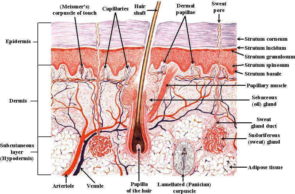
Key facts about the integumentary system; Skin: Functions: chemical and mechanical barrier, biosynthesis, control of body temperature, sensory Layers: Epidermis (Stratum Basale, Spinosum, Granulosum, Lucidum, Corneum) and dermis (papillary, reticular) Mnemonic: British and Spanish Grannies Love Cornflakes Hair: Types: vellus and terminal Structure: Follicle and bulb (shaft, inner root sheath.
